40 picture of the eye with labels
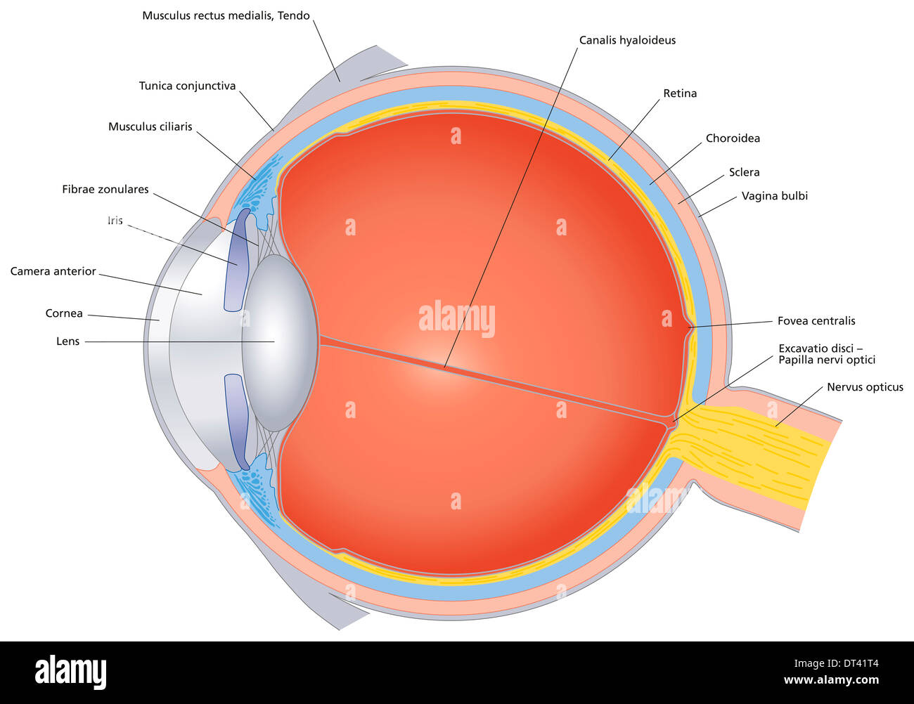
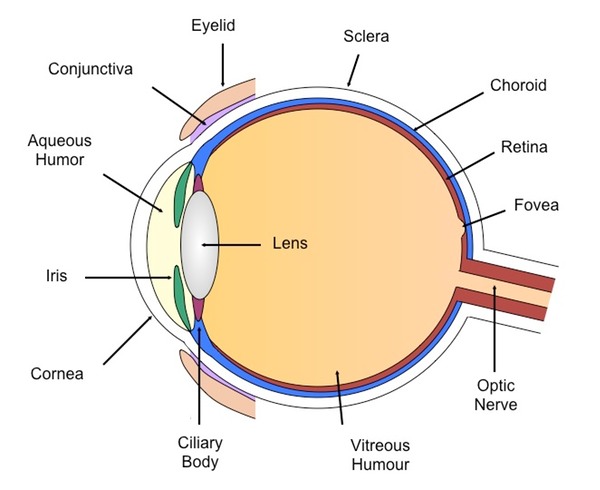
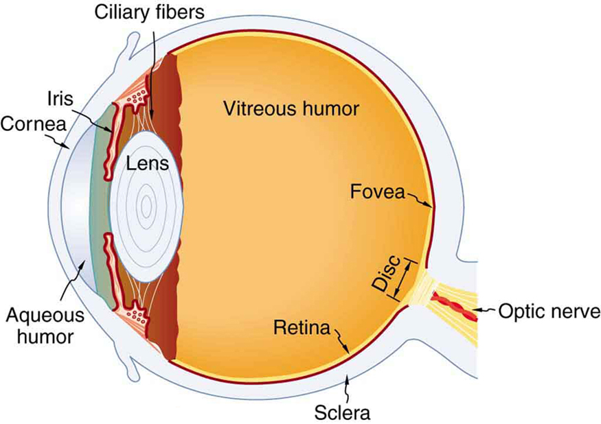
60,892 Human eye anatomy Images, Stock Photos & Vectors - Shutterstock 60,892 human eye anatomy stock photos, vectors, and illustrations are available royalty-free. See human eye anatomy stock video clips Image type Orientation Color People Artists Sort by Popular Biology Healthcare and Medical Icons and Graphics Nutrition human eye anatomy 3d rendering eye visual perception infographic Next of 609 Eye Diagram With Labels and detailed description - BYJUS A brief description of the eye along with a well-labelled diagram is given below for reference. Well-Labelled Diagram of Eye The anterior chamber of the eye is the space between the cornea and the iris and is filled with a lubricating fluid, aqueous humour. The vascular layer of the eye, known as the choroid contains the connective tissue.
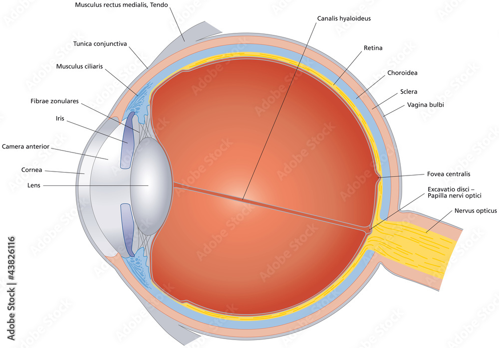
Lens of the Eye - All About Vision The lens of the eye, also called the crystalline lens, is an important part of the eye's anatomy that allows the eye to focus on objects at varying distances. It is located behind the iris and in front of the vitreous body. In its natural state, the lens looks like an elongated sphere — a shape known as ellipsoid — that resembles a deflated ball.

Picture of the eye with labels
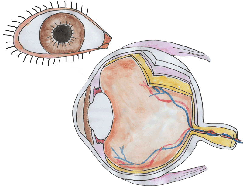
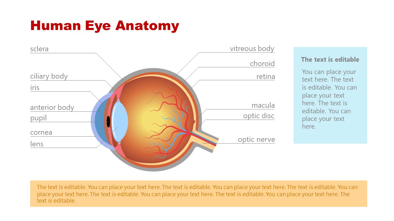
30 Eye-Catching Wine Label Designs For Inspiration The designer used squares to provide the information. 02. The Cloud Factory. The Cloud Factory wine label design looks simple because of its use of two colors only. Yellow and white colors give the label a soft look, which seems to be the intention of the designer and the brand. 03. Label the Eye Worksheet - Teacher-Made Learning Resources - Twinkl In this resource, you'll find a 2-page PDF that is easy to download, print out, and use immediately with your class. The first page is a labelling exercise with two diagrams of the human eye. One is a view from the outside, and the other is a more detailed cross-section. Challenge learners to label the parts of the eye diagram. On the second page, you'll find a set of answers showing ... Anatomy of the Eye | Johns Hopkins Medicine The optic nerve carries signals of light, dark, and colors to a part of the brain called the visual cortex, which assembles the signals into images and produces vision. Posterior chamber. The back part of the eye's interior. Pupil. The opening in the middle of the iris through which light passes to the back of the eye. Retina.
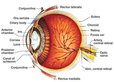
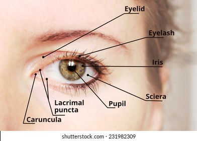
Picture of the eye with labels. Eye Pictures, Anatomy & Diagram | Body Maps - Healthline There are two types: cones make color vision possible, and rods specialize in black-and-white images. Although our eyes can only see in two dimensions, we are able to determine distances and depth... Human Eye Ball Anatomy & Physiology Diagram - eMedicineHealth The orbit is the bony eye socket of the skull. The orbit is formed by the cheekbone, the forehead, the temple, and the side of the nose. The eye is cushioned within the orbit by pads of fat. In addition to the eyeball itself, the orbit contains the muscles that move the eye, blood vessels, and nerves. The orbit also contains the lacrimal gland ... Eye Anatomy: Parts of the Eye and How We See Here is a tour of the eye starting from the outside, going in through the front and working to the back. Eye Anatomy: Parts of the Eye Outside the Eyeball. The eye sits in a protective bony socket called the orbit. Six extraocular muscles in the orbit are attached to the eye. These muscles move the eye up and down, side to side, and rotate the eye. Label the parts of the eye in the picture. The choices are optic nerve ... Find an answer to your question Label the parts of the eye in the picture. The choices are optic nerve, cornea, iris, pupil, lens, and retina. (1) (2) (3) …
Human Eye Anatomy Pictures, Images and Stock Photos Vector illustration of the structure of the eye. Anatomy of the... Structure of the eye, parts of the eye. Retina, macula, blind spot, optic nerve, cones, rods, vitreous humor, ciliary body, lens, pupil, aqueous humor, cornea, iris, sclera, choroid. Houman body parts flat line icons set. Man, woman head, brain... Label Eye Printout - EnchantedLearning.com Label the Eye Diagram. Human Anatomy. Read the definitions, then label the eye anatomy diagram below. Cornea - the clear, dome-shaped tissue covering the front of the eye. Iris - the colored part of the eye - it controls the amount of light that enters the eye by changing the size of the pupil. Lens - a crystalline structure located just behind ... Labelling the eye — Science Learning Hub In this interactive, you can label parts of the human eye. Use your mouse or finger to hover over a box to highlight the part to be named. Drag and drop the text labels onto the boxes next to the eye diagram If you want to redo an answer, click on the box and the answer will go back to the top so you can move it to another box. Human Eye Coloring Page | crayola.com The eye is the organ that collects images and sends them to the brain, so you can see. The eye is protected by the bones of your skull and six muscles. Light comes through the pupil which causes the cornea and lens to focus on an image. When the image is projected through the eye, onto the retina wall, the image appears upside down. Eye ...
31 Most Beautiful Eyes in the World - Woman's World We bet anyone that meets them feels the same way! Getty Images Eyes are naturally beautiful — from the delicate shapes and unique colors to the countless expressions that can be made with them. Blue eyes, brown eyes, green eyes, hazel eyes, gray eyes, and any shade in between are all stunning. Please don't ask us to pick a favorite! 14,721 Eye Drawing Stock Photos - Dreamstime Browse 14,721 professional eye drawing stock photos available royalty-free. Eye. Blue eye drawing on a white background. Drawing: Human body parts, hand, eye and mouth. Colorful drawing: Human body parts, hand, eye and mouth. The eye 3. The eye, part of the human face - hand drawing ancient picture - vinatge postcard, pencil technique. Eye Anatomy Detail Picture Image on MedicineNet.com Picture of Eye Anatomy Detail The eye is our organ of sight. The eye has a number of components which include but are not limited to the cornea, iris, pupil, lens, retina, macula, optic nerve, choroid and vitreous. Cornea: clear front window of the eye that transmits and focuses light into the eye. Anatomy of the eye: Quizzes and diagrams | Kenhub How to learn the parts of the eye. Found within two cavities in the skull known as the orbits, the eyes are surrounded by several supporting structures including muscles, vessels, and nerves.There are 7 bones of the orbit, two groups of muscles (intrinsic ocular and extraocular), three layers to the eyeball… and that's just the beginning. There's a lot to learn, but stay calm!
External anatomy of the human eye (with labels) Solid-Faced Canvas Print
PDF Eye Anatomy Handout - National Institutes of Health of light entering the eye. Lens: The lens is a clear part of the eye behind the iris that helps to focus light, or an image, on the retina. Macula: The macula is the small, sensitive area of the retina that gives central vision. It is located in the center of the retina. Optic nerve: The optic nerve is the largest sensory nerve of the eye.

3d Image Render Of Diagram Of Eye Anatomy With Label For ...
10,401,889 Eyes Images, Stock Photos & Vectors | Shutterstock 10,329,096 eyes stock photos, vectors, and illustrations are available royalty-free. See eyes stock video clips Image type Orientation Color People Artists Sort by Popular Biology Clothing and Accessories Diseases, Viruses, and Disorders Abstract Designs and Shapes human eye evil eye visual perception eye amulet macro photography Next of 103,291
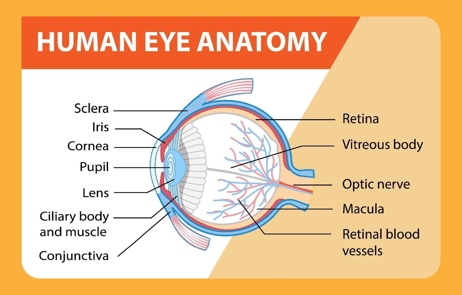
Diagram of human eye anatomy with label 1945551 Vector Art at ...
Label Parts of the Human Eye - University of Dayton Parts of the Eye. Select the correct label for each part of the eye. The image is taken from above the left eye. Click on the Score button to see how you did. Incorrect answers will be marked in red. ...
Eye Histology - Eye (labels) - illustration -
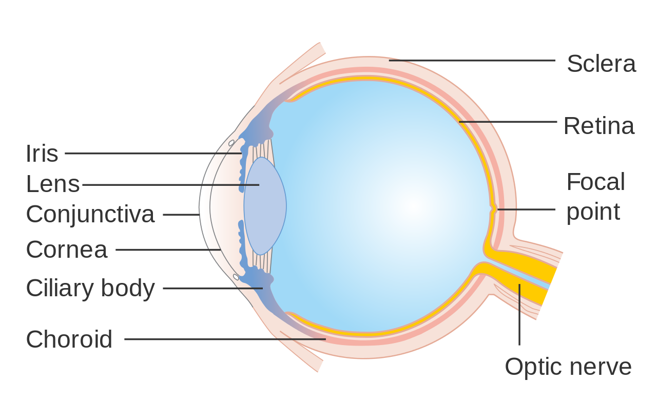
A Picture of the Eye - WebMD Your eye is a slightly asymmetrical globe, about an inch in diameter. The front part (what you see in the mirror) includes: Iris: the colored part; Cornea: a clear dome over the iris; Pupil: the ...

Diagram of the Eye Side View No Labels | Diagram of the eye ...
50,000+ Best Eye Photos · 100% Free Download - Pexels Download and use 50,000+ Eye stock photos for free. Thousands of new images every day Completely Free to Use High-quality videos and images from Pexels. Explore. License. Upload. Upload Join. Eyeball Eyes Eye Close Up Eye Doctor Face Vision Lips Hair Nature Ear Dark Light Sad Look Love Sea. Eye Photos. Photos 55.6K Videos 9.1K Users 551.
13,151 Eye diagram Images, Stock Photos & Vectors | Shutterstock
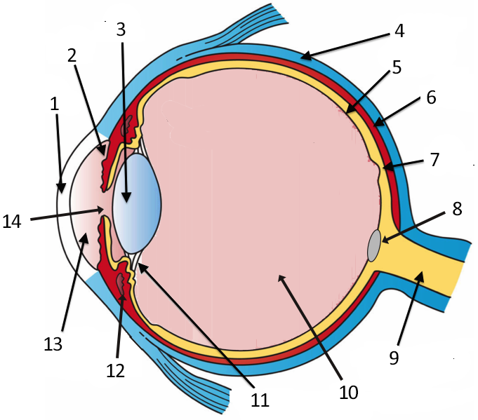
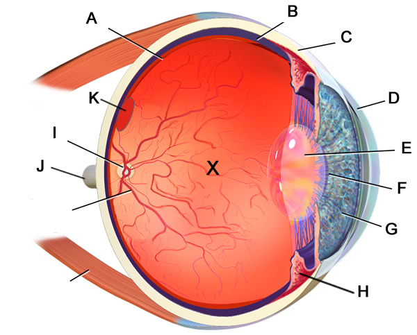
Quiz: Label The Parts Of The Eye - ProProfs Quiz A is pointing to what part of the eye? 2. B is pointing to what part of the eye? 3. C is pointing to what part of the eye? 4. Which part of the eye controls the amount of pressure within the eye? 5. Which part of the eye protects the front of the eye?
Eyes - Layers of Learning
Eye Anatomy Diagram - EnchantedLearning.com Aqueous humor - the clear, watery fluid inside the eye. It provides nutrients to the eye. Astigmatism - a condition in which the lens is warped, causing images not to focus properly on the retina. Binocular vision - the coordinated use of two eyes which gives the ability to see the world in three dimensions - 3D. Cones - cells the in the retina that sense color.

Normal Anatomy of the Eye | Doctor Stock
Eye Anatomy: 16 Parts of the Eye & Their Functions - Vision Center The lens of the eye (or crystalline lens) is the transparent lentil-shaped structure inside your eye. This is the natural lens. It is located behind the iris and to the front of the vitreous humor (vitreous body). The vitreous humor is a clear, colorless, gelatinous mass that fills the gap between the lens and the retina in the eye.

Eye anatomy oblique cross-section - #AN0003
Labelled Diagram of Human Eye, Explanation and Function - VEDANTU The human eye is a part of the sensory nervous system. Labeled Diagram of Human Eye The eyes of all mammals consist of a non-image-forming photosensitive ganglion within the retina which receives light, adjusts the dimensions of the pupil, regulates the availability of melatonin hormones, and also entertains the body clock.
Isolated illustration of the human eye with latin labeling ...
What is an eye mark and why do I need it? - Consolidated Label An 'eye mark' (also known as 'eye spot') is a small rectangular printed area located near the edge of the printed flexible packaging material. A sensor on the form-fill-seal (FFS) machine reads the eye mark to identify packaging material, control the material's position, and coordinate the separation and cutting of the flexible packaging material.

ch 10 labeling eye Diagram | Quizlet
What Does the Eye Look Like? - Diagram of the Eye | Harvard Eye Associates Vitreous Gel: A thick, transparent liquid that fills the center of the eye. It is mostly water and gives the eye its form and shape. Our eyes are vital for seeing the world around us. Keep them healthy by maintaining regular vision exams. Contact Harvard Eye Associates at 949-951-2020 or harvardeye.com to schedule an appointment today.
Eye Anatomy PowerPoint Template - SlideModel
Anatomy of the Eye | Johns Hopkins Medicine The optic nerve carries signals of light, dark, and colors to a part of the brain called the visual cortex, which assembles the signals into images and produces vision. Posterior chamber. The back part of the eye's interior. Pupil. The opening in the middle of the iris through which light passes to the back of the eye. Retina.

DHS - Labeling the Cow Eye Quiz - Quizizz
Label the Eye Worksheet - Teacher-Made Learning Resources - Twinkl In this resource, you'll find a 2-page PDF that is easy to download, print out, and use immediately with your class. The first page is a labelling exercise with two diagrams of the human eye. One is a view from the outside, and the other is a more detailed cross-section. Challenge learners to label the parts of the eye diagram. On the second page, you'll find a set of answers showing ...

Diagram of human eye anatomy with label 1928861 Vector Art at ...
30 Eye-Catching Wine Label Designs For Inspiration The designer used squares to provide the information. 02. The Cloud Factory. The Cloud Factory wine label design looks simple because of its use of two colors only. Yellow and white colors give the label a soft look, which seems to be the intention of the designer and the brand. 03.

Diagram of human eye anatomy with label illustration. | CanStock
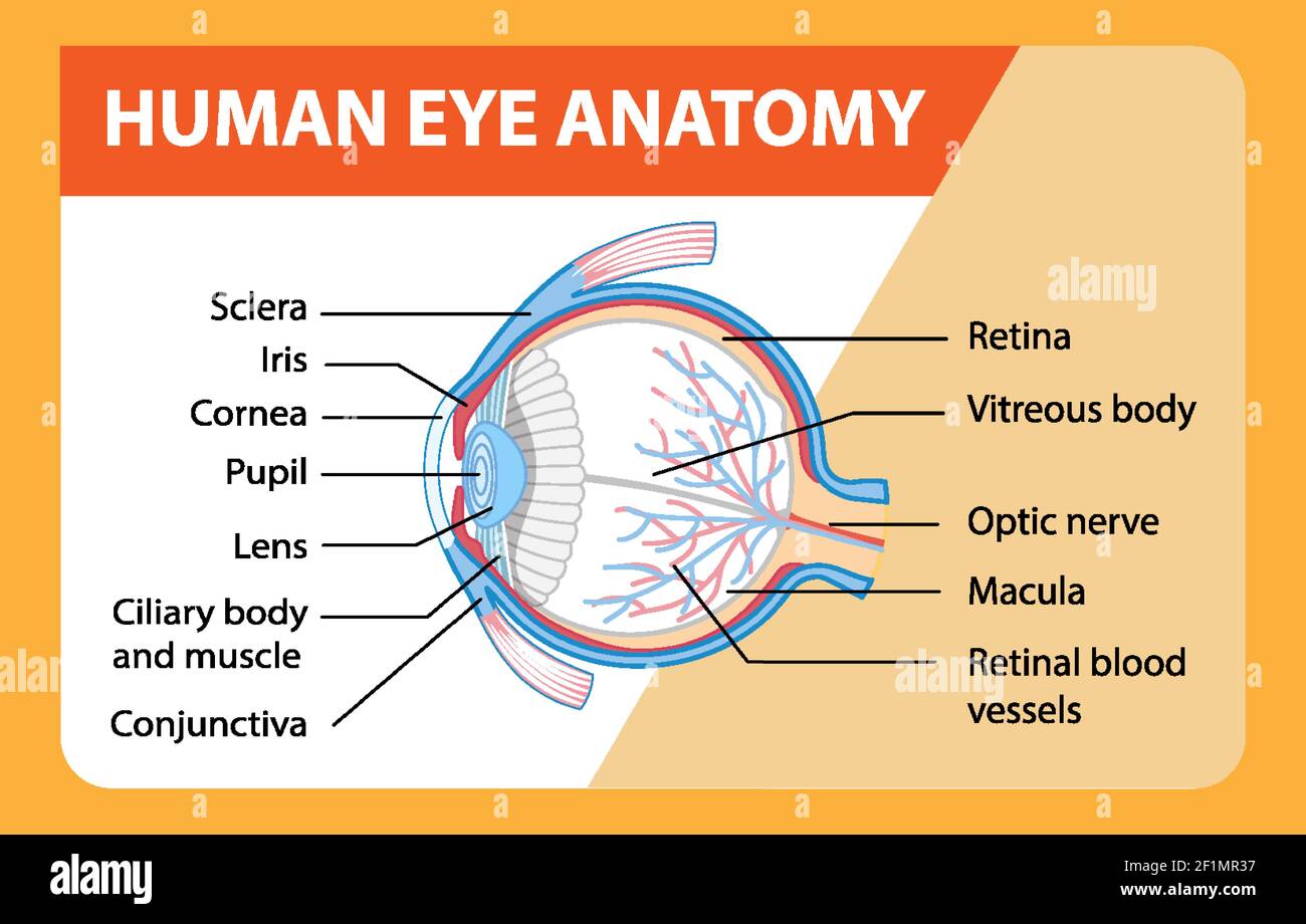
Diagram of human eye anatomy with label illustration Stock ...

Sketch and label V.S. of human eye. - Biology | Shaalaa.com
Label the Eye

Diagram and label the internal structures of the eye, and gi ...
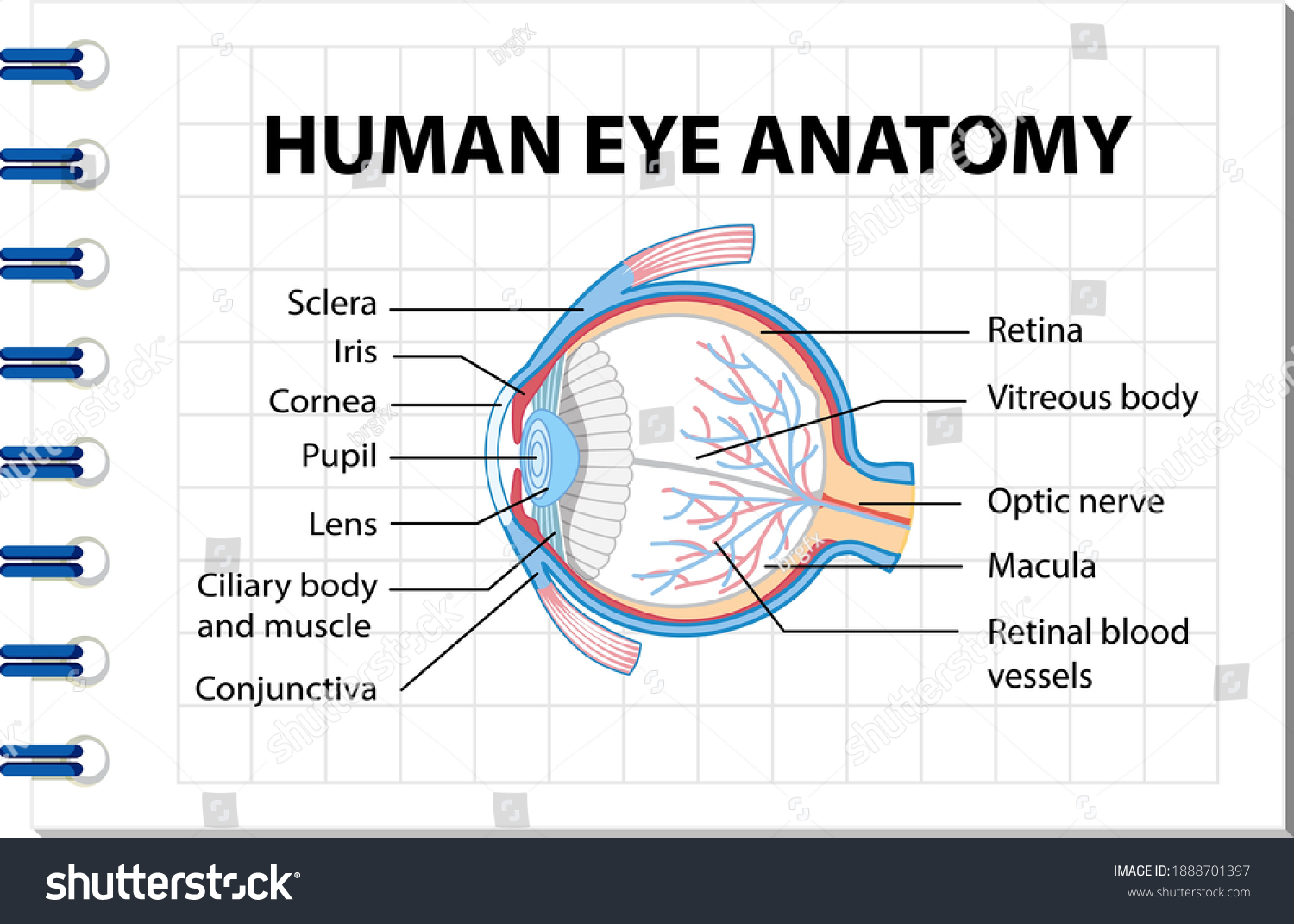
Diagram Human Eye Anatomy Label Illustration Stock Vector ...

How your eye works (parts of the eye)Look After Your Eyes

Labeling human eye** Diagram | Quizlet
Eye Histology - Eye (labels) - Histology illustration -

Anterior (Front) View of the Eye | Doctor Stock

Simple eye diagrams | Easy eye diagram | Labeled eye diagram ...
(249).jpg)
Quiz: Label The Parts Of The Eye - ProProfs Quiz

Diagram human eye anatomy with label Royalty Free Vector
Label the Eye
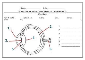
Science worksheets: Label parts of a human eye by Science ...
Anatomy - Vision
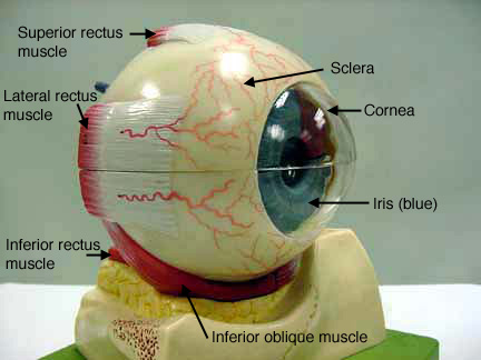
Eye Models
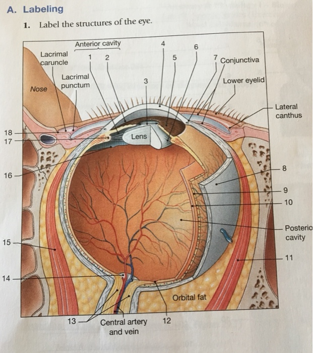
Solved A. Labeling 1. Label the structures of the eye ...
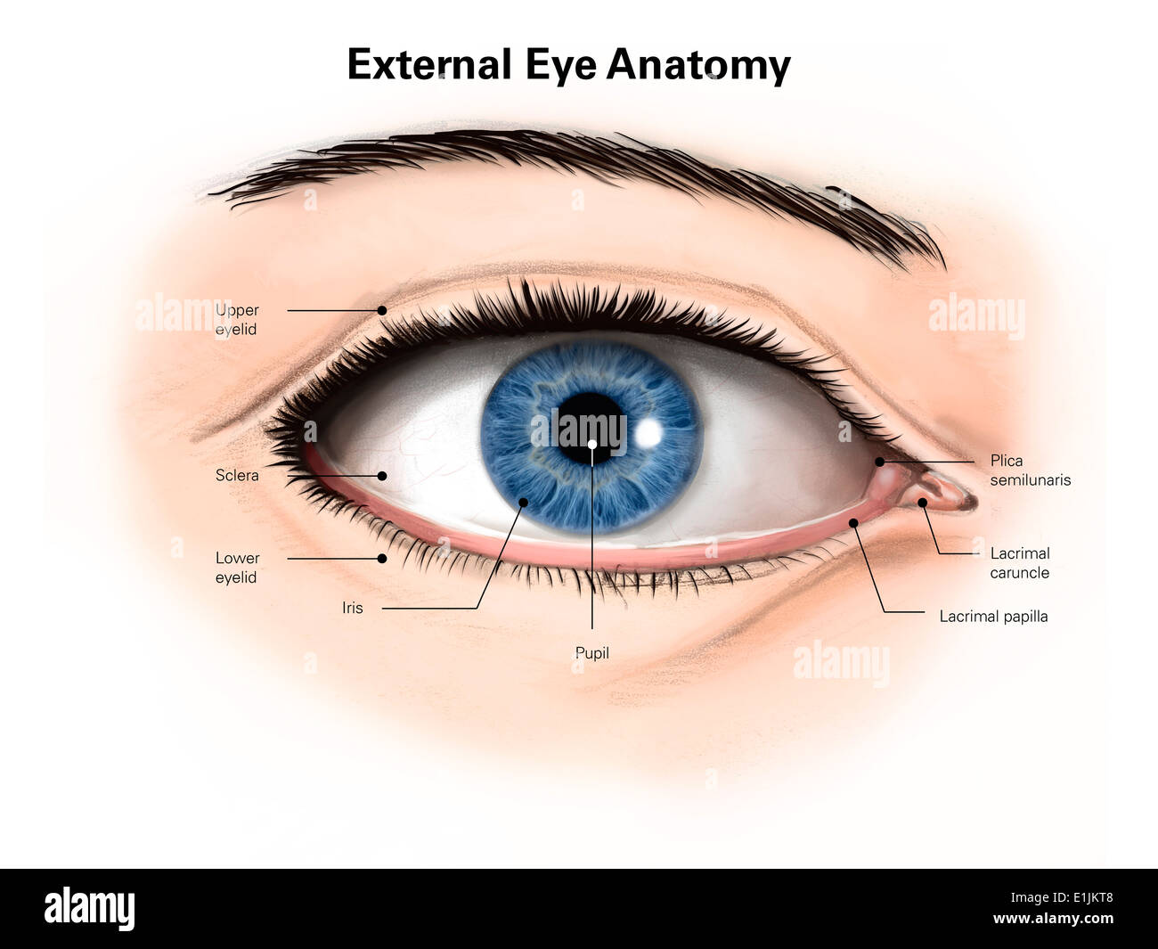
External anatomy of the human eye (with labels Stock Photo ...
5.1 Physics of the Eye and the Lens Equation – Douglas ...

Simple eye diagrams | Easy eye diagram | Labeled eye diagram ...
Structures of human eye with latin labeling. Cross section of ...

Quick Quiz

Complete eye diagram with labels. Courtesy of U.S. National ...
Structure Of Human Eye Without Label Transparent PNG ...
Eye Facts | Cool Kid Facts
Post a Comment for "40 picture of the eye with labels"